Tuesday, November 20, 2018

The Search for Macrofossils within Pete the Peat

by Nick Alex, Lauren Frankel, and Sean Wright

Imagine Grinnell, Iowa 29,000 years before the present (ybp). Instead of seas of corn and soybeans as far as the eye can see, the landscape was comprised of spruce forests and patches of sedge wetlands. While this may sound more like the current day wilderness of Alaska or Canada, cycles of glaciation brought boreal forests south to Iowa. Particularly, the maximum glacial extent in Iowa came from the Des Moines lobe of the Laurentide ice sheet, which covered the north-central region of the state approximately 13,800 ybp (Clayton and Moran 1981). This period of glacial advance followed a spell of minor warming and glacial retreat from 27,000-30,000 ybp (Dyke et al. 2002). Where Grinnell College stands today, about 29,000 ybp one such sedge wetland accumulated plant material in the water only to be uncovered thousands of years later...


Fast forward to the spring of 1960. Grinnell College was undergoing a very familiar process to us: campus construction. The college was building a new fine arts building, the Roberts Theatre. As the Grinnell College students who had the esteemed privilege of witnessing the serene and peaceful Alumni Recitation Hall property get turned into a muddy hole in preparation for the construction of the new Humanities and Social Sciences building know, campus construction has a way of exposing history that has not been seen in ages to the open air (take the Peace Rock for example). This is exactly what happened when the construction of Roberts Theater exposed a deep and extensive peat deposit, tentatively estimated to be 29,000 years old. About 5 meters below surface level, lay a peat deposit, holding the key to understanding the biota of this region prior to anthropogenic alterations to the environment. Peat is a mixture of partially decomposed plant material that has accumulated in a water-saturated environment in the absence of oxygen. In these environments, the greatest plant material accumulates where the temperature is high enough for plants to grow but too low for large amounts of microbial degradation. Peat samples are of particular interest of geologists and palynologists who use the deposits to provide both direct and indirect evidence of interglacial climate and biota.

Picture of the exposed peat during the construction of Roberts Theatre in 1960 (Graham 1962).

One of the ways we can learn more about our peat is to look for evidence of past organic life. This can often be seen as fossils, or in our case macrofossils. Despite the name, these macrofossils need not be large in size but are still expected to be larger than pollen grains. Our aim was to dig through the peat to look for macrofossils such as fragments of vascular plants or bryophytes that could tell us more about the vegetation of the land when the peat was deposited. In the initial work conducted when the peat was discovered, it was primarily pollen and wood fragments “characteristic of northern coniferous forest” that were extracted from the peat (Graham 1962). Because of this, there is more to learn about the plant composition from the examination of macrofossils, including non-woody plants or plants with limited pollen dispersal abilities. 


To examine the peat, we had to decide on a method for first picking apart the portions of peat we were working with, and then further breaking it down to search for macrofossils under dissecting microscopes. The original piece of peat (colloquially called Pete) that we started with was broken down using a rock hammer and a smaller pick in order to split the peat apart and decide pieces that were to undergo further analysis. Once the large pieces of wood were split off and given to the wood group, we placed the smaller pieces of peat into zip sealed bags, eventually to be further broken down in water or potassium hydroxide (KOH) solution and screened through mesh sieves to search for macrofossils under compound microscopes (Faegri et al. 1989, Baker 1996). Once potential macrofossils were found and separated, we placed them on slides for microscopy imaging analysis. We then compared the microscopy images of our macrofossils to previously published and identified macrofossil assemblages.


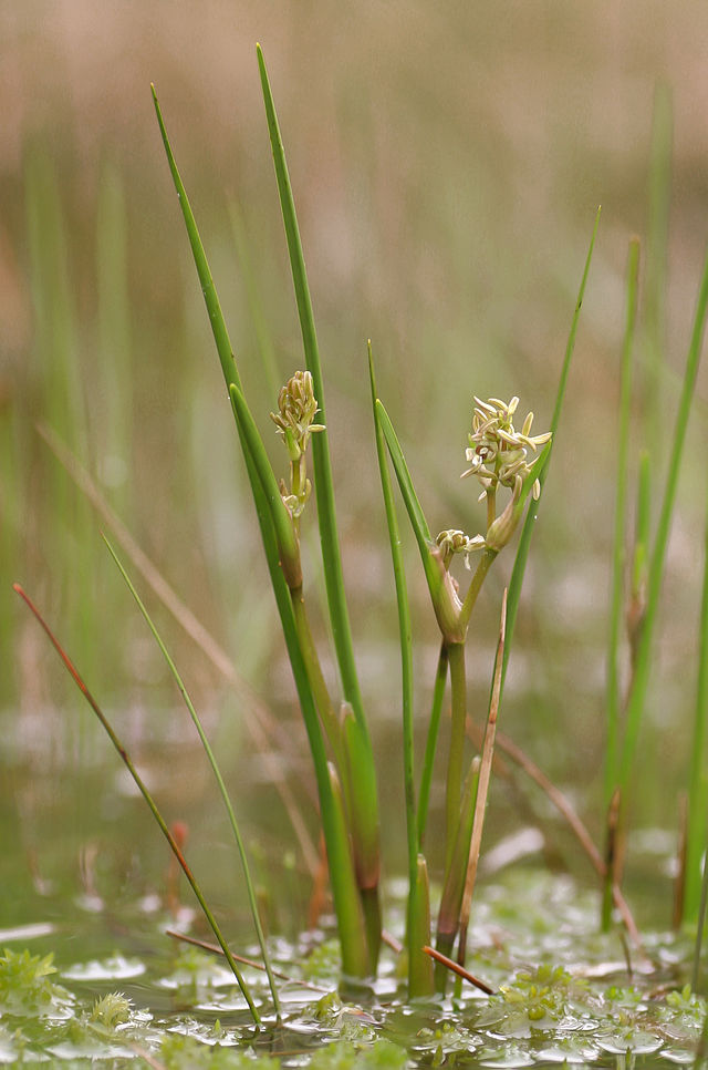
Left: our potential Scheuchzeria palustris rhizome epidermis tissue. Right: Scheuchzeria palustris flowering in the Netherlands (picture from Wikipedia user Bertblok).

We found many macrofossils with an array of rectangular cells about 600 microns long but lacked any indication of stomata, expressed by doughnut-shaped pores used during photosynthesis, leading us to believe this plant tissue was not photosynthesizing. After comparing to Mauquoy & Van Geel 2007, we concluded they were instead rhizome (horizontally growing, underground stem) epidermis from Scheuchzeria palustris, a plant typical of bog and fen waters. At a much lower abundance than S. palustris, we also found plant tissues that displayed some similarity to Betula fusca leaf tissue. However, this sample lacked stomata, as well, and thus were probably stem or root tissue. 


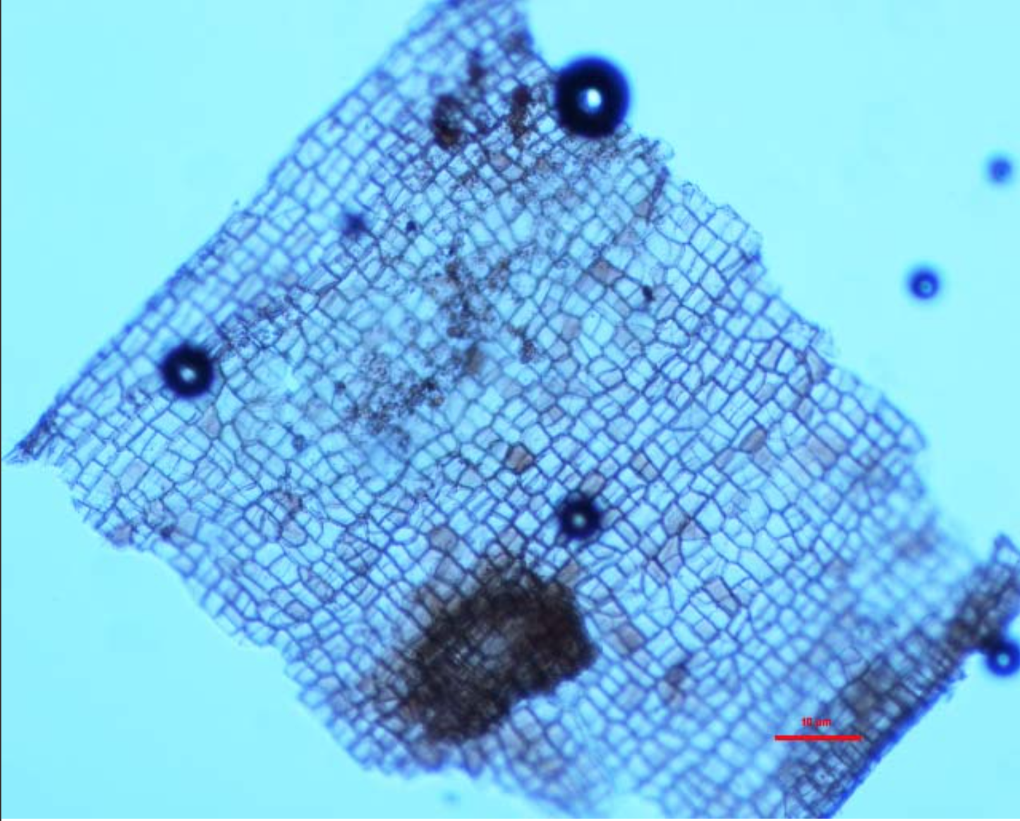
Unidentified stem or root tissue isolated from the Grinnell peat.

These findings lead us to believe that the landscape of Grinnell ~29,000 years ago contained sedge wetlands, the kind of ecosystem conducive to the growth of S. palustris. Sedge wetlands seem like a far cry from the current expansive agricultural operations of corn and soybeans, and grasslands that we see driving around Iowa today. Despite our lack of evidence for a spruce forest, there may have been patches of coniferous forests interspersed with sedge wetlands similar to the composition of Kearney, Nebraska, about 600 km away and also near the southern edge of the Laurentide ice sheet, about 23,000 ybp (Dillon et al. 2018). In the future, an increased reference collection would be of great use to compare macrofossil and wood samples produced from the peat to better understand the paleoenvironment of Grinnell.


Works Cited
Baker, R. G., Bettis III, E. A., Schwert, D. P., Horton, D. G., Chumbley, C. A., Gonzalez, L. A., & Reagan, M. K. (1996). Holocene paleoenvironments of northeast Iowa. Ecological Monographs, 66(2), 203-234.
Clayton, L., & Moran, S. R. (1982). Chronology of late Wisconsinan glaciation in middle North America. Quaternary Science Reviews, 1(1), 55-82.
Dillon, J. S., Stolze, S., & Larsen, A. K. (2018). Late Pleistocene Pollen and Plant Macrofossils from a Buried Wetland Deposit in the Platte River Valley, South-Central Nebraska. Great Plains Research, 28(2), 173-183.
Dyke, A. S., Andrews, J. T., Clark, P. U., England, J. H., Miller, G. H., Shaw, J., & Veillette, J. J. (2002). The Laurentide and Innuitian ice sheets during the last glacial maximum. Quaternary Science Reviews, 21(1), 9-31.
Faegri, K., Kaland, P. E., & Krzywinski, K. (1989). Textbook of pollen analysis (No. Ed. 4). John Wiley & Sons Ltd.
Graham Jr, B. F. (1962). A post-Kansan peat at Grinnell, Iowa: a preliminary report. In Proceedings of the Iowa Academy of Science (Vol. 69, No. 1, pp. 39-44).
Mauquoy, D., & Van Geel, B. (2007). Plant macrofossil methods and studies: mire and peat macros. In Encyclopedia of quaternary science. Elsevier Science.

Monday, November 19, 2018

Unearthing the History of Iowa's Trees

Maggie Loery, Siri Bruhn, and Vishva Nalamalapu

Pop quiz! What is peat?… Not to worry, we didn’t know either until our professor brought a box of it to class, which his colleague had found in the basement of our college's science building. He explained that while peat may look like dirt, it’s actually organic material that has been poorly decomposed due to acidic or anaerobic conditions. It turns out that poor decomposition is good news for us, or for anyone else investigating life at the time the peat was deposited. Since our class, Evolution of the Iowa Flora, is all about understanding how plants in Iowa have changed throughout time, our professor explained that this peat could be a rich source for furthering this understanding.

The box of peat didn’t come with a whole lot of information, but our professor explained that Ben Graham, a former professor at our college, originally found the peat sixty years ago at a construction site on campus (Fig. 1). He published a preliminary report on the peat (Graham 1962) where he mentioned that the peat contained many wood fragments preserved in "cellulosic form," and identified some wood fragments as Tamarack (Larix larcina). He estimated the peat was deposited between 12,000 and 130,000 years ago, corresponding to the time between the early Wisconsin and Illinoian glacial stages.


Figure 1. Note in the box of peat

Once we selected a 24x16 cm chunk of peat to study, we fondly named it “Peter,” and gently began disaggregating it. We uncovered fragments of wood, as well as fragments of leaves and insects. Though we hoped to find pollen as Ben Graham did, we were sadly not able to. Thus, we split the investigation into three groups: Team Wood, Team Macrofossils, and Team Reference. While we, as Team Wood, tackled the identification of wood fragments, Team Macrofossils did the same for other plant fragments. Team Reference set out on a scavenger hunt to collect reference wood fragments from trees on campus. Our hope was if they visualized their reference samples with the same techniques as we used on our samples, we could compare the two and better identify the historic wood fragments.

We began by separating cells. Our professor explained that we could create a maceration fluid as described by Huang and Yeung (2015), which would separate the cells, allowing us to search for particular cell types and features that distinguish between trees. We had to macerate the samples for longer than expected as the components binding the cells were not adequately digested. And that was not the only complication we encountered. Preparing microscope slides involved carefully transferring small quantities of cells onto a block of gel on the slide, melting the gel, and then placing a coverslip on top (Fig. 2)… Easier said than done. The cells were often either not adequately digested or over-digested, and the slides often had bubbles that made the cells difficult to visualize. We should probably admit that we never did perfect this slide-preparation technique, but we did manage to make slides for seven of our samples that allowed us to view the features we were hoping to see.


Figure 2. Microscope slide preparation setup

Our identification relied on three cellular characteristics: bordered pits, helical thickenings, and the presence of vessel cells (Fig. 3). Bordered pits, which resemble donuts, but are far too small to eat, are abundant in gymnosperm tracheid cells. Helical thickenings are spiral ridges in tracheid and vessel cells. Angiosperms have small bordered pits compared to gymnosperms. Using the findings of Baker et al. on the composition of the flora of Iowa during the Holocene, we used the size of bordered pits to distinguish between three trees that grew in Iowa around the time the peat was likely deposited: larch, pine, and spruce (Baker et al. 1990). Spruce cells have bordered pits in a single row, pine cells have larger bordered pits in a single row, and larch cells have bordered pits in pairs. Vessel cells, which comprise long, water-conducting structures, are only present in angiosperms.


Figure 3. Our key to identify wood fragments

Keeping an eye out for these features, we visualized our microscope slides (Fig. 4). The wood fragments seemed to include larch, pine, spruce, and an angiosperm! We observed single rows of ~20 µm diameter bordered pits in the tracheids of three samples, leading us to believe these wood fragments were from spruce trees. Single rows of ~30 µm bordered pits of another sample led us to believe this wood fragment was from a pine tree. Pairs of bordered pits in tracheids of another sample indicated this wood fragment was from a larch tree (Fig. 5). We only found helical thickenings in one sample. They appeared to be on vessel cells and were dotted by bordered pits (~50 µm diameter), which indicated this wood fragment was from an angiosperm (Fig. 6).


Figure 4. Maggie and Siri visualizing microscope slides.


Although we originally intended to compare our images to Team Reference’s, many of their images showed structures we did not observe (i.e. bordered pit fields in vessel elements; Fig. 7). We were, however, able to see that the bordered pits on the pine tree reference (~50 µm diameter) were slightly larger than those on ours (~30 µm diameter), which could indicate mis-identification, or could simply represent an actual range of sizes.


Figure 7. Team Reference’s bordered pit field in a vessel element

Soon, we’ll have better information about the age of our peat too. In addition to our wood identification, we prepared and submitted three wood fragments to Beta Analytic for radiocarbon dating. We’ll let you know once we have a better idea of when these larch, pine, spruce, and angiosperm trees were inhabiting what is now Grinnell College!

So, in our two short weeks of analyzing “Peter’s” wood fragments, we found evidence that the Grinnell flora included larch, pine, spruce, and angiosperm trees when the peat was deposited. If you want to see what Iowa was like back then, drive a few hours up to northern Minnesota and imagine you’re instead in Iowa long ago. Compare the landscape you see to Iowa today, and consider the evolution from conifer woods to prairie to the acres on acres of corn and soybeans you see today. Studies like ours and that of Baker and colleagues allow us to understand how and when these changes took place. Our work is just the beginning; there is plenty of other work to be done, and there’s plenty of peat left to do it!


Works Cited
Baker, R. G., Chumbley, C. A., Witinok, P.M., & Kim, H. K. (1990). Holocene Vegetational Changes
in Eastern Iowa. Journal of the Iowa Academy of Science, 97, 167-177.

Hoadley, R. Bruce. (1990). Identifying Wood: Accurate results with simple tools. Newtown, CT: Taunton Press.

Graham, B. F. (1962). A Post-Kansan Peat at Grinnell, Iowa: A Preliminary Report. Proceedings of the Iowa Academy of Science, 69, 39-44.

Yeung, E. C. T., Stasolla, C., Sumner, M. J., & Huang, B. Q. (2015). Plant microtechniques and protocols. Springer International Publishing.

Sunday, November 18, 2018

References for Pete

A Short Investigation of a (Very) Long Paleobotanic History

Andrea Baumgartel, Emily Burgess, Isaac Ferber

Paleobotany is the study of historic plant communities and species using information gathered from preserved geological records of past environments. It’s important because it tells us about what our environment used to be like, like how plant morphology changes over time, how old a species is, and how ecological communities undergo change over time. Paleobotany can also give us clues about what we can expect as our climate changes in the current day by telling us what the ecological communities of an area were like in the past, and what the environment was like back then. It’s common for communities of plants, including many of Iowa’s own, to have cyclical patterns of population and distribution decline, and recovery in response to similar shifts in climate, among other factors. Paleobotanic studies can be done using fossilized plants or pieces of plants that have escaped decomposition in peat bogs, and still remain relatively intact after thousands of years.
            Several studies have been completed in Iowa using paleobotany to reconstruct past environments. In 1996 Baker et al. did an analysis of the floodplain alluvium (material deposited by a river over years of flooding) of Roberts Creek in northeastern Iowa to determine how the plant community surrounding the floodplain had changed over the last 11,000 years. They found that there was a succession of boreal forest, followed by deciduous forest, prairie, and oak savanna, leading in the modern agricultural area. This is a good example of how paleobotany can inform us on what the environments we live in used to look like, in terms of plants at least.
            During the construction of the Robert’s Theatre on Grinnell College Campus, a layer of peat was uncovered during excavation, and a preliminary analysis of what was preserved in the peat was done by Ben Graham in 1962, who found some wood fragments and pollen. The rest he left for us, and in this year’s Evolution of the Iowa Flora class we set out to see what else we could find and identify.
Our investigation didn’t involve the actual unearthing (or rather, un-peating) of the identity of the unknown materials in the peat (which we’ve affectionately named “Pete”) itself, but rather the crafting and imaging of already-known specimens (dubbed “reference materials”) that could potentially be, or be related to, said unknowns. Once the ancient unknown organic matter (e.g., small-to-microscopic wood bits, leaves, stems, pollen, etc.) is extracted from the peat, the reference materials can be used comparatively in order to help determine the identification of those unknowns.
While there are certainly a fair amount of available reference images existing online, it’s always advantageous to make one’s own reference materials, because 1) it helps to have consistent imaging methods and settings (i.e., the prep & imaging processes will be the same for both references and unknowns) and 2) you can never 100% guarantee that the reference identifications you find online are actually what they say they are. Also, we’ve collected reference materials from the actual location (Grinnell Iowa) that had the potential to be found in the peat, and thus the reference materials we find will be more directly relevant to this location’s past.
The reference materials in question are wood samples from the following tree genera, all located throughout the central most parts of Grinnell College’s campus: Alnus glutinosa (alder), Betula nigra (birch), Carpinus caroliniana (hornbeam), Fraxinus americana (ash), Pinus strobus (pine), Populus deltoides (cottonwood), and Tsuga caroliniana  (hemlock). Our imaging began with pollen samples collected earlier by professor Vince Eckhart, which ended up not being used, as numerous attempts to detect pollen within the actual peat itself were unsuccessful. However, we remain optimistic that pollen evidence will be found from the peat sample in the future, as the seminal paper by Ben Graham included notes verifying that the peat did in fact contain pollen, despite our findings.
With no ancient pollen to identify, our focus moved from creating pollen references (of which there is a fantastic online database anyway), and we shifted our goals to collecting wood from around campus, processing them in the same way as the peat fragments, and imaging it so the rest of the class could use our slides to identify their mystery wood. The first task was to search campus to collect samples from the modern equivalent of trees that might be in the peat. After a day of collecting, we broke down our samples, which were branch fragments, into small bits that fit into small vials. These were soaked in 5% potassium hydroxide (KOH) to break down the wood in the same way the peat was processed. After a week in the solution at a constant 45°C, we were ready to begin to make our slides for imaging. Just putting chunks of wood on a glass plate wasn’t going to cut it, so we needed to dye and divide the samples into pieces as small as possible, ideally to expose individual cells.
We didn’t find definitive literature on how to process specifically macerated wood samples into something microscope friendly, so we spent a day working to create our own method. The system that yielded the best results involved two slide plates, one for staining the wood cells with safranin (aka basic red 2), and the other for cleaning excess stain and cutting the wood into the smallest pieces possible in deionized (distilled) water. The newly stained wood was cleaned in deionized water, and after waiting for the water to evaporate, the sample was sealed in melted gelatin under a cover slide. Our best samples were permanently enclosed with clear nail polish. We imaged cells we identified as containing visible, important structures, specifically searching for vessel elements in angiosperms, along with helical structures, and border pits in gymnosperms (Figures 1 & 2). These parts were the most likely to be identifiable in the ancient wood collected from the other groups.


Fig 1. Modern Alnus glutinosa sample with visible vessel elements and perforation plate.


Fig 2. Modern Carpinus caroliniana sample with visible helical structures within a vessel element.


References

Graham, B. F. Jr. 1962. A Post-Kansan Peat at Grinnell, Iowa: A Preliminary Report. Proceedings of the Iowa Academy of Science 69: 39-44.


Baker, R.G., E. A. Bettis III; D. P. Schwert; D. G. Horton; C. A. Chumbley; L. A. Gonzalez; M. K. Reagan. 1996. Holocene Paleoenvironments of Northeast Iowa. Ecological Monographs 66: 203-234.


The Grinnell Peat: Preserved; Uncovered; Forgotten; Re-discovered; Explored.


For an undetermined number of years (not exceeding 58), a re-purposed cardboard box sat in the basement of the Noyce Science Center. Finding and opening the box, my faculty colleague Andrew Graham ("Graham A," whose position is in Chemistry and Environmental Studies) found: (1) several brick-sized clumps of dry soil, rich in organic matter, rather casually protected with heavy paper; (2) handfuls of sneeze-provoking dust; and (3) a copy of a 1962 scientific article written by beloved Grinnell Biology Professor Ben Graham (1920-2009; "Graham B"). 

In the article Graham B told the origin story of the box's contents. The construction of Grinnell College's Roberts Theatre in 1960 uncovered a 50 cm layer of soggy, compressed peat, 5 m below the ground surface. Peat deposits form in places like bogs, where acidity, lack of oxygen, and low temperatures hinder decomposition. In this way, active areas of peat formation represent "sinks" for carbon, while ancient deposits represent important sources of fossils. Recognizing that the deeply buried "Grinnell Peat" could reveal prehistoric plant communities, Graham B and his students sampled the peat. Graham (1962) was "preliminary report" of their findings, including a broad age estimate (300,000 - 20,000 years ago) and notes on the identities of fossil wood and pollen the peat contained. 

We don't yet know what fraction of the material Graham B and his students examined ended up in the mysterious, forgotten box, nor do we know when the packaging took place. We do think that the preliminary report had no sequel. Graham A graciously transferred the box to my care, recognizing my department's claim and imagining that my students might learn by attempting to expand Graham B's 1962 report. 

BIO 305 (Evolution of the Iowa Flora) includes a unit on the history of Iowa vegetation. Past students' research into that history, however, mainly consisted of field trips to sand prairies and other sites that reveal some of that history, and of spotlighting what other scientists had discovered. Blog posts from 2013 and 2015 document some of this work. In 2018 my BIO 305 students made some of their own discoveries, extracting and attempting to identify fossils from the Grinnell Peat, and making reference collections of contemporary plants for comparison. In the posts that follow, teams of students reveal some of their findings and what it took to discover them. Meanwhile, I pledge not to hide their source material in a mystery box in the basement.