Monday, October 30, 2023

Urban Birds


Consequences of Urban Landscapes on Wintering Bird Communities

Oscar Angell, Erik Hallberg, Solveig Olson

Grinnell College BIO 368

With rapidly increasing rates of urbanization happening worldwide, scientists have sought to understand how human environmental sprawl affects other organisms. There are many different species whose numbers have been negatively impacted as a result of increased signs of human presence such as habitat loss and litter we find in our oceans. However, there is not much information available on how overwintering bird populations are affected as a result of human presence as most studies have been carried out during the breeding season. There is knowledge of synanthropic bird species—such as pigeons, house sparrows, and crows—that benefit from human interaction. This apparent variation in birds’ responses to urbanization presents a certain nuance to the question: “How are overwintering bird populations affected by urban landscapes?”. An article published in 2016 set out to answer this question, considering how urban habitat structure, as well as human effects such as pollution, noise, and artificial light, impact overwintering bird communities in southern Poland.

As Ciach et al. point out, “urban areas account for almost 3% of the Earth’s land surface, and this proportion is continually rising” (547). The increasing expansion of human environments is one of the most harmful threats to non-human organisms, and vast species extinction on a global scale continues as a result of urban sprawl and its destruction of crucial natural areas. It is therefore necessary that the scientific community examines the effects of urbanization on biodiversity. One group of organisms that is largely present in urban areas is birds, and the specifics of their interactions and community assemblages within urban landscapes has yet to be studied in depth. Ciach et al. aimed to fill this gap in knowledge by looking at the ways in which bird communities wintering in urban areas are impacted by the habitat traits of an urban environment, including habitat structure, food availability, and light and noise pollution. To investigate this critical question, they used a line transect method to measure the composition, density, and interseasonal similarity of bird assemblage within 56 sample plots chosen at random in Kraków in 2014-15. Notably, Kraków has an urbanization gradient, making it an especially useful study site.

Ciach et al.’s main findings are that bird populations and species diversity are negatively impacted by noise pollution and building density, and are positively impacted by food availability, urban greenery, and in some cases, light pollution. They hypothesized that noise pollution decreases bird population and diversity by drowning out bird calls. These bird calls are required for many bird behaviors, such as group foraging, coordination, and flocking. With some species unable to complete these behaviors, the bird diversity and population decrease. High building density was also found to decrease bird diversity. The authors hypothesized that this was due to a lack of shelter limiting the bird species that can thrive in urban areas. Conversely, greenery coverage (farmland or urban greenery) was found to increase the diversity of bird types, as well as the overall number of birds. The hypothesis offered for greenery’s positive impact is that it increased the amount of shelter, allowing for more species to survive. Unsurprisingly, food availability also played a role in determining population density. In urban areas, populations of synanthropic bird species were significantly higher in areas with large amounts of human byproducts that the birds would eat. The final and most surprising finding was that light pollution may have a positive impact on bird populations. Ciach et al. observed that as a result of light pollution, birds would start foraging behaviors earlier in the mornings, and keep foraging later into the night although this was not true in all situations, and some species were negatively impacted by light pollution. However, the researchers found the overall population did better with light pollution.

Table 8. Environmental predictors of winter bird community traits


 As an extension of the work of Ciach et al., our group studied bird communities on a college campus in rural Iowa, a space of approximately 120 acres surrounded by the town of Grinnell and vast expanses of agricultural crop farming. Similar to the methods of Ciach et al.’s study, we used a walking line transect, relying on the Merlin ID app to record and identify the presence of bird species with call recognition. For three weeks in October, we collected data from three different sections of the college campus, each with different combinations of habitat structure and greenery. Consistent with the findings of Ciach et al., bird species richness—reflected by the number of species counted in each location—increased with a higher proportion of urban greenery and building cover. While our study is far more limited in its temporal and spatial span, its findings are relevant and complementary to those of Ciach et al. and expand on their work by examining a much smaller and less densely populated urban landscape: a college campus. Considering future implications of our work, we believe that Merlin ID is a helpful tool for examining the effects of urbanization on bird communities on other college campuses.

Sources Cited

Ciach, M., Fröhlich, A. (2017). Habitat type, food resources, noise and light pollution explain the species composition, abundance and stability of a winter bird assemblage in an urban environment. Urban Ecosystems 20, 547–559. https://doi.org/10.1007/s11252-016-0613-6

Hanski, I. (2011). Habitat Loss, the Dynamics of Biodiversity, and a Perspective on Conservation. AMBIO 40, 248–255. https://doi.org/10.1007/s13280-011-0147-3

Lincoln, S., Andrews, B., Birchenough, S., Chowdhury, P., Engelhard, G., Harrod, O., Pinnegar, J., & Townhill, B. (2022). Marine litter and climate change: Inextricably connected threats to the world's oceans. Science of The Total Environment, 837, 155709. https://doi.org/10.1016/j.scitotenv.2022.155709

Monday, November 14, 2022

A Hitchhiker (Pollen)’s Guide to the Galaxy (Iowa Flora)

Pollen. It makes you sneeze, fertilizes plants, and transports you through time to ancient landscapes. Wait, what was that about time travel? Well, maybe we can’t quite hop onto a pollen grain like a microscopic version of Doc Brown’s DeLorean, but it can play a compelling role in paleobotany, the study of fossilized plant remains. With the help of two of our own enthusiastic Docs– Dr. Eckhart of the 2020s and Dr. Graham of the 1960s– and a very old chunk of partially decomposed plant matter (peat), we embark on an adventure through time in hopes of catching a glimpse of the flora of our college’s campus… 27,000 years ago.

If you haven’t explored the other entries on The Natural History of Grinnell College, allow us to catch you up. We are undergraduate students in Grinnell, IA taking a course entitled Evolution of the Iowa Flora under the instruction of Vince Eckhart. In the 1960s, a professor by the name of Ben Graham stashed away a sample of peat that was unearthed during the construction of Roberts Theater. Picking apart this peat, Graham and his students began to decipher the clues of Iowa of Old. Mysteriously, Graham didn’t seem to follow up as he proposed in his 1962 paper. Neither did anyone else, until 2018. Just a few years ago, the peat was unearthed once again, that time from a cardboard box in the science building basement. After a COVID hiatus from in-person classes, we (along with Team Macrofossil and Team Wood) pick up where the 2018 students left off, this time with revised methods. To behold this peat, to view its pollen, insect exoskeletons, papery plant tissue, and wood under the microscope is to face deep history. These fragments of Iowa flora lived long before climatic changes gave rise to the most well-known ecosystem in our area– the prairie– about 10,000 YBP. Today, as we walk over a lawn manicured by the College in a state where about 85% of the land is used for agriculture, we wonder what other remnants of the past might reside just a few meters below our feet. We begin to wonder what it was like here when the organic matter was laid down on the floor of a boreal sphagnum bog (like those in modern-day Canada) 27,000 years ago. Written here is our contribution to the tale of the Grinnell peat, a story barely known but slowly revealing itself to those who are curious enough to look closely.


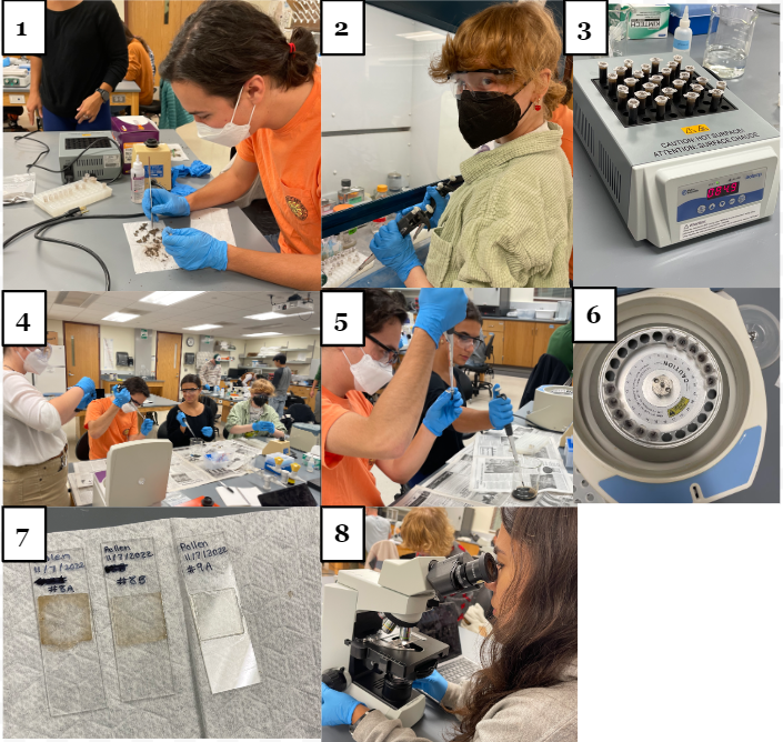
As scientists in a biology laboratory, our portal to the past takes the form of microscope slides. We prepared 30 microscope slides as outlined in Figure 1. In November 2022, we examined and photographed the slides. We compared our photos taken at 200x magnification with the images on the Global Pollen Project website (https://globalpollenproject.org/Taxon). Using this technique, we identified pollen grains from the genera of Pinus (pine), Picea (spruce), and Quercus (oak). 


Previously, Ben Graham identified the pollen of “spruce, fir, pine, alder, maple, numerous 

‘betulaceous’ grains, and others characteristic of northern coniferous forest, or transition thereto”. Thus, our findings corroborate Graham’s identification of spruces but also evidence that there were oaks present on the land now referred to as the Grinnell College campus. Our identification time was limited, and thus suggest further examination of our slides to identify other pollen grains present– whether they corroborate Graham’s other findings or introduce new evidence. 


Figure 1. Picture collage of the steps in the preparation of pollen for analysis. 


We used modified and simplified methods based on a newly published protocol developed in 2022, by Santos and Lerdu. This procedure differs from previous methods of pollen extraction, as it doesn’t use corrosive acids. We created 26 microcentrifuge tubes, each filled with crushed peat dirt. Using a vortex mixer and microcentrifuge, we mixed the peat sample with potassium hydroxide to dissolve impurities, rinsed a number of times using distilled water, and used zinc chloride to create a density gradient that suspended the pollen in the liquid. Using distilled water again, we centrifuged the solutions, making the pollen clump into a pellet. We added glycerine to each tube to give the pollen something to stick to and made microscope slides for examination. For more detailed methods, see Santos and Lerdu 2022, or additional note at the end of this post.


A note on contamination: A classmate of ours on Team Macrofossil brought to our attention a grain of pollen reminiscent of a fidget spinner that he found while exploring macrofossils under the scope. We identified it as belonging to Oenothera, the evening primroses. However, this finding does not necessarily mean that there were evening primroses present in the Grinnell flora 27,000 years ago. It
could, rather, be evidence of present-day flora in the Grinnell College biology corridor: We suspect this pollen to be contamination brought into our lab from our professor’s
Clarkia xantiana (an evening primrose) research laboratory just down the hall. 


During our analysis, we observed that the most common type of pollen in our sample was made of a single pollen grain with two air bladders, arranged similarly to Mickey Mouse ears. While the pollen of many conifers share this general shape, we determined that these grains were most likely from a pine tree based on its small size. Notably, spruce pollen, which we identified as being the second most abundant pollen in the sample, has a very similar grain shape to pine, with similar function, so a critical difference is their relative sizes; Picea grains are roughly 110 to 150 micrometers long compared to Pinus grains, which are 50 to 110 micrometers long (Figure 2 and 3). The quantity of the pollen suggests that pine trees in particular were abundant around Grinnell when this peat formed, though we may not be able to know exactly how close they were to the site of our peat. The air bladders we see in both Pinus and Picea suggest the pollen was mostly spread by the wind rather than animal pollinators, as research has shown that these structures improve the pollen’s ability to pollinate by wind dispersal, acting almost like a parachute, slowing the pollen’s fall and allowing to travel further. As a result, the pollen can travel massive distances under the right conditions, making it hard for us to assume where exactly the pine and spruce may have been in relation to the peat.


Figure 2: Pinus (pine) pollen sourced from Grinnell peat and photographed under 200x magnification. At right is a 2018 reference slide prepared by Professor Eckhart with a slightly different procedure, leading to the color variation between the two. The figure on the left, sourced from Leopold & Zaborac-Reed (2014), was critical in helping distinguish Picea and Pinus based on size and shape characteristics.


Figure 3: Picea (spruce) pollen photographed under 200x magnification. At left is the spruce reference slide prepared by Professor Eckhart in 2018. 


As we continued our analysis we found a less abundant pollen grain that we believe is Oak (Figure 4). The shape and size of the pollen convinced us that this is a match, and tells us that, similar to present-day Grinnell, oak trees were here 27,000 years ago.


Figure 4: Quercus (oak) pollen taken using a microscope set to 200x magnification. The image on the left is a reference slide made in 2018, showing contemporary oak pollen.
The image on the right is of oak pollen found in the peat.

Amidst the pine, spruce, and oak pollen grains, we saw a rather abundant pollen grain we didn’t recognize (Figure 5). As we worked on identifying this sample, we learned that as pollen dries, it might change shape. We suspect that the grains we photographed are on the drier side, which made identification more of a challenge. A 1972 paper by V. Sh. Vagababian describes the morphology of magnolia family pollen. Vagababian measured the length of these pollen grains to be around 50 μm, which is similar to the size of our samples. If this is a match, tulip poplar trees may have been present in Grinnell’s ancient landscape! 


Figure 5: The two images on the left are pulled from the page on PalDat, a palynological database, for Liriodendron tulipifera (commonly known as tulip poplar) showing a dry pollen grain on the top and a hydrated grain on the bottom. At right, the pollen we suspect to be tulip poplar under 200x magnification. Does it look like a match?


In collaboration with the wood and macrofossil group, we hope to increase our knowledge about Grinnell’s flora. However, due to time constraints and unknown finds, we know there is still so much left to do to uncover more knowledge about our college campus’ past. As you traverse our campus or wherever you are, remember that you are walking on a profound mass of history– what other stories does the land have to tell us? Stay curious. 


Authors:

Sonia Edassery, Joanie Fieser, Athena Frasca, Isabelle Jacqmotte-Parks, and Sam Takahashi


Notes: 


The following procedure describes the collage in Figure 1, stepwise: 

In a sterile environment, we created 26 microcentrifuge tubes with about 0.2 mL of mashed peat dirt and 1.5 mL distilled H2O (1). We then vortexed and spun the solution at 3000 rpm for 4 mins. We then added 1.5 mL 10% KOH to only the pellet and vortexed (2). Next, the tubes were put into an 85°C dry bath for 6 mins (3). We then vortexed the solution and spun it at 3000 rpm for 4 mins. The resulting solution looked dark and opaque, like coffee. We added 1.5 mL distilled water to only the pellet, vortexed it briefly, and centrifuged the tubes at 3000 rpm for 4 minutes (4). This step was done 6 times until the solution was light, transparent brown. We then removed the supernatant, or extra liquid, and air-dried the leftovers (containing pollen!) for 5 mins.1 mL of 1.9 g/mL zinc chloride was then added and vortexed (5) and spun at 1000 rpm for 4 min. We then transferred the supernatant to new microcentrifuge tubes with 1.5 mL distilled water, then vortexed and spun them at 3000 rpm for 4 minutes, twice (6). An equal amount of pellet, containing the pollen, and 80% glycerine was finally added to the tube. We made microscope slides by placing a drop of tacky glue on a clean microscope slide with some contents of the tube (7). We used microscopes set to 200x magnification, meaning every 10 units measured to be 45 micrometers (8).


References:

Graham Jr, B. F. (1962). A post-Kansan peat at Grinnell, Iowa: a preliminary report. In Proceedings of the Iowa Academy of Science (Vol. 69, No. 1, pp. 39-44).


Leopold, Estella & Zaborac-Reed, Stephanie. (2014). Biogeographic History of Abies bracteata (D. Don) Poit. in the Western United States. 


Rudney de Almeida Santos & Marie-Pierre Ledru (2022) Acid-free protocol for extracting pollen from Quaternary sediments, Palynology, 46:1, 1-8, DOI: 10.1080/01916122.2021.1960916


V. Sh. Agababian (1972) Pollen Morphology of the Family Magnoliaceae, Grana, 12:3, 166-176, DOI: 10.1080/00173137209429874


“Don’t Drink the Dirty Chai” - An Examination of Macrofossils in the Grinnell College Peat Deposit

 Isabella Vergara, Hayden Bhavsar, Anna Lipari, Nate L’Esperance

Driving down Interstate 80, or almost anywhere else in Iowa, what do you see? It’s often fields, sometimes rolling, with sparse groups of deciduous trees poking out from the endless expanse of corn and soybeans. To most, the lack of, well, anything at all, creates a bland impression of the Hawkeye State. However, what if we were to tell you it wasn’t always this brown slab of corn? Would you ever assume that at some point, Iowa was a coniferous swampland akin to the vast forests of the United States’ Northwoods regions or Western Ontario? Grinnell Professor Ben Graham has helped to prove that Iowa indeed has an interesting, decidedly very different past.

Way back in the ancient, bygone, prehistoric era of 1960, Professor Ben Graham began to inspect the excavation site of what would eventually become a new Fine Arts center for Grinnell College in Grinnell, Iowa. After digging a mere 2.5 feet below the excavation floor, he began to find a 3 foot thick layer of dark brown, and loose organic matter. Graham found this layer to have an abnormally high water content of 82.4% at a depth of 16 feet, relative to 26% above to 28% below this layer (Graham, 1962.) Later, this layer was dated to 26-27,000 years before present. The layer consisted of peat, a type of organic soil resulting from prehistoric wetlands (Graham, 1962.) Peat originates from acidic freshwater bogs. Because of the acidic, low-oxygen conditions, most dead organic matter that fell into the water became preserved, as the waters would not decompose the debris (Xintu, 2009.) As time passed and more earth filled in over bogs, the resulting preserved organic material hardened into peat deposits. 


Due to the lack of decomposition, Ben Graham found that his excavated peat samples were full of preserved bits of leaves, seeds, stems, and pollen from many prehistoric plants. Formally, these preserved bits are called plant macrofossils. These macrofossils are classified as “remains large enough to be visible without a microscope” (NOAA.) We set out with the objective of indiscriminately extracting these plant macrofossils from the peat samples. What we find in the peat samples may give us more insight into what kind of plants may have existed in place of Grinnell College many tens of thousands of years ago. Similar extractions of the peat have yielded interesting results in 1962 and 2018, which we hope to build on with new techniques that may allow us to find more macrofossils.


As a class, we decided to pick one chunk of peat to share among team pollen, team wood, and us, team macrofossil. We placed small lumps of our peat into beakers with either purified water or KOH (an acidic solution intended to remove compounds that might interfere with imaging) and warmed them to dissolve the chunks into a coffee-colored sludge that we fondly described as a dirty chai


Figure 1: Left - the “Dirty Chai” consisting of broken up peat samples. We are testing deionized water and an acidic solution, KOH, for breaking up samples. Right - Anna and Nate searching through the samples for macrofossils with bronze fine mesh sieves (106 and 250um, shown in the bottom right corner). Macrofossils were obtained with tweezers and prepared on microscope slides.


Then, we gently sieved through the sludge under a microscope, to look for any little bits of plant material we could see. If we happened upon anything that looked plant-like, we used a pair of tweezers to gently place the plant bits on slides with a drop of tacky glue, so we could visualize them under our scopes later (Mauquoy et al., 2010). Once our tacky glue had dried, we took pictures of each of our slides and tried to describe and identify the plant material we were seeing based on published macrofossil keys (Lévesque et al., 1988). Then, we were able to compare our findings to the slides that students from previous years had made. 


In 2018, team macrofossil found what they thought was rhizome epidermis from the bog plant Scheuchzeria palustris. Our collection was much different from the 2018 team’s. We found a variety of plant parts amongst the peat. While we were unable to use these pieces to figure out with certainty what kinds of plants grew in the area since they were so small, we made informed guesses about the types of tissue we unearthed. Many of the macrofossils we found were wood fragments that required maceration for identification, a process which our group was not equipped to complete. Despite the abundance of unidentifiable wood, we found a higher volume of plant tissue compared to team Macrofossil of 2018. It’s possible that our fragment of peat had a higher concentration of plant material than the other, or that we tended to isolate smaller fragments than the other team, as there may be differences in our sampling and searching methods despite our efforts to replicate their work. 


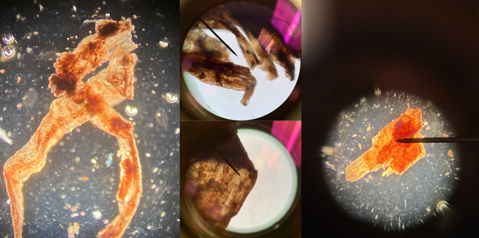
In addition to wood, we found charcoal pieces (courtesy of Team Wood) which suggest the presence of fire in the peat bog. We also found significant amounts of epidermis tissue from unknown plants (figures 3 and 4). These samples tended to be very small, without distinguishing characteristics that pointed towards a specific species. We saw two main categories, tissue with short, square-ish cells and tissue with more elongated cells. The lack of stomata, donut shaped cellular holes that allow for gasses to move in and out of the plant, suggests that much of this tissue is bark or root tissue. We did find a few samples which we believe to be leaf tissue with stomata (see figure 4). Other interesting finds include a fossil resembling an amoeba (figure 5), a partial winged seed (figure 5), and what might be some sort of grass sheath (figure 1). 


Figure 2: A ring-shaped tissue fragment possibly from a grass sheath of Scheuchzeria palustris. The elongated cells and shape ring are vaguely similar to leaf scars on S. palustris. Left: 40x, Right: 400x magnification. 


Figure 3: Pieces of fibrous plant tissue with elongated cells. It is possible these come from some kind of marsh grass. 40x magnification. 


Figure 4: Plant tissue with short cells. The bottom right image shows tissue with stomata, donut shaped holes that allow for gasses to move in and out of the plant (shown in the bottom right corner at 400x). This piece probably came from a leaf or stem since these are the areas where stomata are present. Left image and top right at 40x magnification. 


Figure 5. Odds and ends. Left - Tissue fragment, possibly a piece of a winged or wind-dispersed seed. 40x magnification. Top right - a piece of charcoal found by Team Wood. 40x magnification  Bottom right - potentially a testate amoeba, evidenced by what appears to be a shell. 400x magnification.


Though we can’t draw any strong conclusions, our macrofossils hint at changes in prehistoric plant life. Our peat was formed around 27,000 years ago, but we don’t know the range of time covered. Team macrofossils in 2018 might have found different fragments because they extracted macrofossils from a piece of peat that formed when the area was a different kind of environment. Maybe they were looking at a piece from when it was a fen while our piece was from when it was a bog. In this case our findings might show environmental change. For instance, the presence of charcoal in our peat shows evidence of fire. And while we have been unable to make any genus-level identifications, this does say something about the diversity of plant life that once survived in this area– a far cry from the endless fields of corn that seem to dominate today’s Iowa, and a hint at the multiplicity of species that still exist today in the gaps between. 


Works Cited 


Lévesque, P.E.M., Dinel, H., Larouche, A. (1988). Guide to the identification of plant macrofossils in Canadian peatlands. Canadian Government Publishing Centre. 


Mauquoy, D., Hughes, P., van Geel, B. (2010). A protocol for plant macrofossil analysis of peat deposits. Mires and Peat 7(6), 1-5. 


National Oceanographic and Atmospheric Administration (NOAA). Plant Macrofossil. National Centers for Environmental Information.


Xintu, Liu. (2009). Conditions of Peat Formation. Coal, Oil, Shale, Natural Bitumen, Heavy Oil and Peat - Vol II. Encyclopedia of Life Support Systems (EOLSS)